Dr. Tavares has training in dentistry and expertise in the field of developmental biology, with emphasis in cardiovascular and musculoskeletal craniofacial biology and development. During his MS. and Ph.D. studies, he received broad training in anatomy, histology, and cell and molecular biology. For the execution of his Ph.D. dissertation, he received a highly competitive grant award from the Brazilian government to travel abroad and work in the laboratory of Dr. Raymond Runyan at the University of Arizona where he later became a postdoctoral research scholar. Because of his training in dentistry, Dr Tavares started a second post-doctoral fellowship in the laboratory of Dr. David Clouthier at the University of Colorado Anschutz Medical Campus. During that time, he studied normal and abnormal craniofacial development by working with different transgenic mouse lines, zebrafish, and different cell lines relevant to craniofacial development. Following this time at the University of Colorado, Dr. Tavares became an Assistant Research Professor at the George Washington University (GW) where he received an R03 grant application from NIH to fund his independent research. At GW, Dr Tavares analyzed development of the cranial bones, jaws and ear using transgenic mice and Xenopus in order to study the role of SIX1 and SIX1-associated genes in neural crest cell patterning and differentiation, and in the development of musculoskeletal elements of the face and skull. In vitro cell culture complements the work with animal models.
Dr. Tavares believes that the work with mouse, Xenopus and cell culture provides a rich research environment for trainees at different levels (e.g., undergraduate and graduate students, postdoctoral scholars). By uncovering the role of SIX1 during craniofacial development, these studies will help patients and their families understand the genetic causes of SIX1-related birth defects (e.g., branchio-oto-renal syndrome, craniosynostosis), and lead to preventive and corrective strategies in the near future.
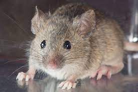
Mammalian model
Mus musculus (mouse)